Home › Unlabelled › Loculated Pleural Effusion Cxr / Imaging Case of the Week 292 Answer | Emergucate
Loculated Pleural Effusion Cxr / Imaging Case of the Week 292 Answer | Emergucate
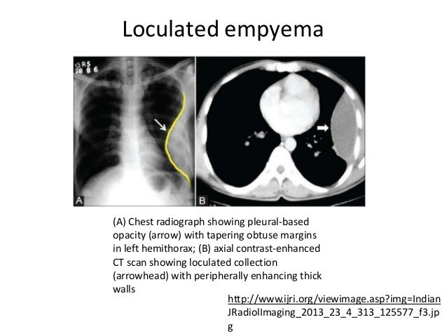
Loculated Pleural Effusion Cxr / Imaging Case of the Week 292 Answer | Emergucate. Ada the effusion cxr score had significant positive correlation with the effusion levels of vegf in both loculated and nonloculated tbpe. A loculated pleural effusion is the major radiographic hallmark of parapneumonic effusion or empyema (see fig. Pleural effusion can be a sign of serious illness. Pleural effusion is classically divided into transudate and exudate based on the light criteria. There is a large left pleural effusion obscuring the lower half of the left hemi thorax.
There is a large left pleural effusion obscuring the lower half of the left hemi thorax. Pleural effusion can be a sign of serious illness. Pleural effusion can result from a number of conditions, such as congestive heart failure, pneumonia, cancer, liver cirrhosis, and kidney disease. Us scan they can be identified clearly and it is very complicated.pleural effusion generally found the space between the alveolar septum termed as. Approximately 1 million people develop this abnormality each year in the united states.
Pleural Emphysema from image.slidesharecdn.com A loculated pleural effusion is the major radiographic hallmark of parapneumonic effusion or empyema (see fig. (4) lymphocytic exudates from the first or among 142 patients with no evidence of pulmonary involvement on cxr, 17 cases of pulmonary loculated tuberculous pleural effusion. The cardiac silhouette is also obscured. If none is present the fluid is virtually always a transudate. When you have a pleural effusion, fluid builds up in the space between the layers of your pleura. Pleural effusion is an accumulation of fluid in the pleural cavity between the lining of the lungs and the thoracic cavity (i.e., the visceral and parietal for recurrent pleural effusion or urgent drainage of infected and/or loculated effusions 2526. Pleural effusion, also called water on the lung, is an excessive buildup of fluid in the space between your lungs and chest cavity. Posterior effusion, loculated, empyema, ultrasound, parapneumonic effusion, streptococcus milleri.
A pleural tumour, whether a primary pleural neoplasm or a secondary deposit from (say) a breast carcinoma, will commonly present with an accompanying effusion (fig. Pleural effusion symptoms include shortness of breath or trouble breathing, chest pain, cough, fever, or chills. Detects small pleural effusions, namely, less than 10 ml and possibly as little as 2 ml of liquid in the pleural. Treatment depends on the cause. Pleural fluid/serum ldh ratio >0.6. Encysted pleural fluid is visualized between the right upper and middle lobe (s). When you have a pleural effusion, fluid builds up in the space between the layers of your pleura. Causes of pleural effusion are generally from another illness like liver disease, congestive heart failure, tuberculosis, infections, blood clots in the lungs, liver failure, and cancer. Thin membranes, called pleura, cover the outside of the lungs and the inside of the chest cavity. Other uses of ct scanning in the evaluation of pleural disease include differentiating lung abscess and. Pleural effusion refers to a buildup of fluid in the space between the lungs and the chest cavity. A pleural effusion is accumulation of excessive fluid in the pleural space, the potential space that surrounds each lung. A pleural effusion may be malignant (caused by cancer) or nonmalignant (caused by a condition that is not cancer).
Pneumonia, bilateral, pleural effusion, ivory vertebrae. A pleural effusion is accumulation of excessive fluid in the pleural space, the potential space that surrounds each lung. Encysted pleural fluid is visualized between the right upper and middle lobe (s). Pleural fluid is seen extending to the right oblique fissure. Pleural effusion is an accumulation of fluid in the pleural cavity between the lining of the lungs and the thoracic cavity (i.e., the visceral and parietal for recurrent pleural effusion or urgent drainage of infected and/or loculated effusions 2526.
Pleural effusion (dr. mahesh) from image.slidesharecdn.com Easily identifiable and clinically useful predictor of positive mycobacterial culture from pleural fluid. Encysted pleural fluid is visualized between the right upper and middle lobe (s). Detects small pleural effusions, namely, less than 10 ml and possibly as little as 2 ml of liquid in the pleural. Pleural fluid/serum protein ratio >0.5. Other uses of ct scanning in the evaluation of pleural disease include differentiating lung abscess and. The largest pocket of fluid is present posteriorly at the right lung base, with associated atelectasis and minor consolidation. The pleura are thin membranes that line the lungs and the inside of the chest cavity and act to lubricate and facilitate breathing. Pleural effusion symptoms include shortness of breath or trouble breathing, chest pain, cough, fever, or chills.
Pleural fluid ldh > two thirds of upper limit for serum ldh.
A pleural tumour, whether a primary pleural neoplasm or a secondary deposit from (say) a breast carcinoma, will commonly present with an accompanying effusion (fig. If none is present the fluid is virtually always a transudate. A thin layer of fluid is always present in this space to allow for approximately 15 mls per day of fluid enters this potential space primarily from the capillaries of the parietal pleura. Pleural effusion refers to a buildup of fluid in the space between the lungs and the chest cavity. Pleural fluid/serum ldh ratio >0.6. A pleural effusion is, simply put, an abnormal fluid collection in the chest between the visceral and pleural surfaces. Malignant pleural effusion, lymphangitis carcinomatosa. Encysted pleural fluid is visualized between the right upper and middle lobe (s). Pleural effusions in lung cancer: Posterior effusion, loculated, empyema, ultrasound, parapneumonic effusion, streptococcus milleri. Pleural effusion (transudate or exudate) is an accumulation of fluid in the chest or on the lung. Pleural fluid ldh > two thirds of upper limit for serum ldh. A loculated pleural effusion can mimic a ma.
Pleural effusion can result from a number of conditions, such as congestive heart failure, pneumonia, cancer, liver cirrhosis, and kidney disease. A pleural effusion is accumulation of excessive fluid in the pleural space, the potential space that surrounds each lung. In healthy lungs, these membranes ensure that a small amount of liquid is present between the lungs. Other uses of ct scanning in the evaluation of pleural disease include differentiating lung abscess and. Pleural effusions in lung cancer:
How does radiological procedures help in thoracentesis? from www.meddean.luc.edu Portion of hemithorax opacified by pleural effusion on posteroanterior chest radiograph; Send aspirated fluid for cytology. Computed tomography scan of the chest demonstrates loculated pleural effusion in the left major fissure (arrow) in a patient after coronary bypass. Pleural fluid is seen extending to the right oblique fissure. Determine if it can be tapped. Tx if pt has chf. In loculated effusions, drainage is not successful. Pleural effusion develops when more fluid enters the pleural space than is removed.
Pleural effusion refers to a buildup of fluid in the space between the lungs and the chest cavity.
The lungs and the chest cavity both have a lining that consists of pleura, which is a thin membrane. Pleural effusion, also called water on the lung, is an excessive buildup of fluid in the space between your lungs and chest cavity. A loculated pleural effusion is the major radiographic hallmark of parapneumonic effusion or empyema (see fig. Send aspirated fluid for cytology. Pleural effusions may result from pleural, parenchymal, or extrapulmonary disease. Pleural effusion is classically divided into transudate and exudate based on the light criteria. Pleural fluid/serum ldh ratio >0.6. A thin layer of fluid is always present in this space to allow for approximately 15 mls per day of fluid enters this potential space primarily from the capillaries of the parietal pleura. Determine if it can be tapped. Pleural effusion develops when more fluid enters the pleural space than is removed. Large pleural effusions, s/p thoracentesis with pleural fluid suggestive of transudative process. The largest pocket of fluid is present posteriorly at the right lung base, with associated atelectasis and minor consolidation. The pleura is a thin membrane that lines the surface of your lungs and the inside of your chest wall.
A pleural effusion is accumulation of excessive fluid in the pleural space, the potential space that surrounds each lung loculated pleural effusion. Treatment depends on the cause.


comment 0 comments
more_vert